Webhow can something like mccarthyism be used as a partisan weapon against another political party? The Creative Commons Public Domain Dedication waiver (http://creativecommons.org/publicdomain/zero/1.0/) applies to the data made available in this article, unless otherwise stated in a credit line to the data. In current practice, the use of extracellular contrast agents usually allows to determine the diagnosis of most focal liver lesions and should be favored as first imaging approach for the characterization of focal liver lesions and as baseline and follow-up imaging in oncologic patients. In our study, 11(78.6%) of 14 cases were asymptomatic and the other 3 cases found the neoplasm occasionally or during the treatment of other diseases.
For instance, An et al. a single abnormal collection of blood vessels that is less than about 1.5 inches (about 4 centimeters) Abdom Imaging 35:337345, Vernuccio F, Cannella R, Meyer M et al (2019) LI-RADS: diagnostic performance of hepatobiliary phase hypointensity and major imaging features of LR-3 and LR-4 lesions measuring 1019 mm with arterial phase hyperenhancement. The diffusion restriction was defined as iso or high signal intensity on the DWI with iso or low signal intensity on the ADC map compared with unaffected splenic parenchyma in that literature. [15]. Sureka J & Jakkani R. Clinico-Radiological Spectrum of Bilateral Temporal Lobe Hyperintensity: A Retrospective Review. Gadobenate dimeglumine-enhanced MRI in (a) arterial phase and (b) hepatobiliary phase demonstrates two liver metastases with different signal characteristics. The funding bodies played no role in the design of the study and collection, analysis, interpretation of data, and in writing the manuscript. 2B Large regenerative nodules. All Rights Reserved. A 65-year-old patient with HCV-related cirrhosis and hepatocellular carcinoma. Int J Surg Case Rep. 2012;3(10):492500. The etiology is unclear, but may result from different mechanisms, including developmental and acquired causes. https://doi.org/10.1007/s00330-020-06687-y, Tsuboyama T, Onishi H, Kim T et al (2010) Hepatocellular carcinoma: hepatocyte-selective enhancement at gadoxetic acid-enhanced MR imagingcorrelation with expression of sinusoidal and canalicular transporters and bile accumulation. They offer high-quality diagnostic services that enable the treatments. PET-CT scanned 60min and 120 to 150min after FDG administration. Decreased expression of OATP1B3 is one of the steps of hepatocarcinogenesis and leads to HBP hypointensity [75, 76]. Google Scholar. The MRI hyperintensity is the white spots that highlight the problematic regions in the brain. In addition, with DWI and ADC findings on MRI, the diagnosis will be more reliable. Med (Baltim). However, the hyperintensity area appears a little lighter comparatively. 3 patients underwent PET-CT. Activation of -catenin protein causes uncontrolled hepatocyte proliferation and overexpression of OATP1B3 responsible for iso- or hyperintensity on HBP [32, 45, 46]. With contrast-enhanced CT or gadolinium-enhanced MRI, the lesions show nodular enhancement at the periphery, followed by centripetal infilling over the next 510 min.
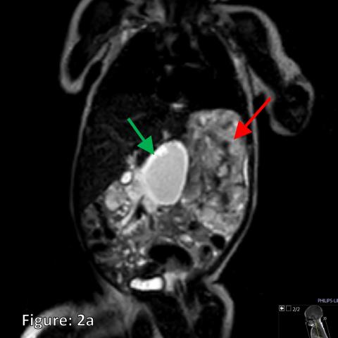
Left column: a uptake in hepatocytes and (b) corresponding signal characteristics in normal liver. J Magn Reson Imaging 36:686696, Kitao A, Matsui O, Yoneda N et al (2018) Differentiation between hepatocellular carcinoma showing hyperintensity on the hepatobiliary phase of gadoxetic acid-enhanced MRI and focal nodular hyperplasia by CT and MRI. 38. Intralesional calcification was not seen in all 5 lesions. Eur J Radiol 92:110, Park HJ, Kim YK, Park MJ, Lee WJ (2013) Small intrahepatic mass-forming cholangiocarcinoma: target sign on diffusion-weighted imaging for differentiation from hepatocellular carcinoma. J Hepatol 61:10801087, Kitao A, Zen Y, Matsui O et al (2010) Hepatocellular carcinoma: signal intensity at gadoxetic acid-enhanced MR imagingcorrelation with molecular transporters and histopathologic features. [1] It was reported as an asymptomatic lesion with slight female predominance. Chen, NX., Wang, ML., Wang, HX. Cystic change or necrosis was absent in all 12 patients. Apart from the progressive spoke-wheel enhancing pattern, DWI and ADC findings will add value to the diagnosis of SANT. Three types of blood vessels are observed including cord capillaries, splenic sinusoids, and small veins. Patients were not required to give informed consent to the study because the analysis used anonymous clinical data that were obtained after each patient agreed to treatment by written consent. Foregut cysts usually possess a definable enhancing wall.[3]. J Am Coll Radiol 14:S314S325, de Ridder J, de Wilt JH, Simmer F et al (2016) Incidence and origin of histologically confirmed liver metastases: an explorative case-study of 23,154 patients. WebLesions were located in the left hepatic lobe in 13 cases, in the right lobe in 11, and in the caudate lobe in 2. Specifically, -cateninactivated HCAs, -cateninactivated-inflammatory HCA and HCAHCC are expected to show a hyperintense signal on HBP, and HCAHCC might show a faster sinusoidal excretion because of their increased MRP3 expression [44, 46]. 180 Fenglin Road, Xuhui District, Shanghai, 200032, China, Department of Pathology, Zhongshan Hospital, Fudan University, No. Richard C Semelka,* Diego R Martin,† and N Cem Balci‡ *Department of Radiology, University of North Carolina, School of Medicine, Chapel Hill, North Carolina †Department of Radiology, Emory University Hospital, Atlanta, Georgia, USA ‡University of Kocaeli, Derince, Kocaeli, Turkey. CD8 and CD86 positive expression was presented in 6 and 7 cases respectively. 34 Zhongshan Bei Road, Licheng District, Quanzhou, Fujian, China, Department of Radiology, Zhongshan Hospital, Fudan University, No. symmetric hyperintensities within the pons, substantia nigra, medulla, anterior horns of the spinal cord, and ventral nerve roots 3. . WebLesions were located in the left hepatic lobe in 13 cases, in the right lobe in 11, and in the caudate lobe in 2. In addition, practitioners associate it with cerebrovascular disorders and other similar risks. The lesion shows continued progressive enhancement on delayed phase(1I). The largest lesions are confluent in the right lobe, showing hypointensity on unenhanced T1-weighted images (a), centrifugal enhancement from arterial (b) to portal (c) and transitional (d) phase, hyperintensity on T2-weighted (e) images. Diffusion weighted imaging in the liver. Sclerosing angiomatoid nodular transformation of the spleen: CT and MRI features with pathologic correlation. Recently, Yoneda et al. The use of hepatobiliary contrast agents is particularly important in the evaluation of liver metastases because it increases sensitivity for detection of metastases compared to CT or extracellular agents providing high tumor-to-lesion contrast on HBP [66, 67]. The likelihood of these observations depends on the patient's on age, gender and risk factors such as oral contraceptives, steroids, history of glycogenosis [10,11,12,13,14,15,16,17]. In the United States, you can find a network of imaging centers that facilitate patients. Springer Nature. The maximum standardized uptake value (SUVmax) was 4.5, 5.1, and 3.8 respectively. Webt2 hyperintense lesion in the right hepatic lobeknox blox for dogs. On T2WI, 10 cases(83.3%)showed hypointensity and 2 cases (16.7%) showed hyperintensity with central hypointensity.
However, sometimes SANT with iron deposition can cause a susceptibility artifact showing hypointensity on both DWI and ADC map, with a signal decrease on in-phase image. In addition to the above considerations and prior to any decision on patient management, it is important to investigate whether the patient has any prior cross-sectional imaging available and to compare all prior examinations, particularly the oldest available one, with the current examination, in order to assess for lesion stability in size or changes of imaging presentations over time. Falk GA, Nooli NP, Morris-Stiff G, Plesec TP, Rosenblatt S. Sclerosing Angiomatoid Nodular Transformation (SANT) of the spleen: case report and review of the literature. Top row: 53-year-old woman with breast cancer and focal nodular hyperplasia. 2010;16(13):156776. Although FNH may increase in size in 315% of cases, these lesions do not evolve to malignancy and their management is conservative [27, 28]. Insights Imaging 12, 8 (2021). Periventricular white matter hyperintensities, Suppose you are having a medical issue, and your physician recommends an MRI. PLoS ONE 8:e70896. Eur Radiol 22:642653, Article C Concurrently performed MRI demonstrates the lesions to be T2 hyperintense and D avidly enhancing in the arterial phase. On CT, all 5 lesions showed hypodensity on non-contrast images (Figs. However, it also exists in young and middle-aged people who have a history of other medical issues. Eur J Radiol 81:38773882, Asayama Y, Tajima T, Nishie A et al (2011) Uptake of Gd-EOB-DTPA by hepatocellular carcinoma: radiologic-pathologic correlation with special reference to bile production. [16] A more significant signal decrease could be seen on DWI and T2WI because of a more significant susceptibility effect of DWI. Am J Surg Pathol 36:16911699, Reizine E, Amaddeo G, Pigneur F et al (2018) Quantitative correlation between uptake of Gd-BOPTA on hepatobiliary phase and tumor molecular features in patients with benign hepatocellular lesions. Dr. Paxton Daniel answered 12 patients underwent MR scan. The vast majority of focal liver lesions are hyperintense on T2-weighted magnetic resonance (MR) images. Google Scholar, Vernuccio F, Dioguardi Burgio M, Barbiera F et al (2019) CT and MR imaging of chemotherapy-induced hepatopathy. One patient complained of right upper quadrant pain, two patients discovered the lesions incidentally during routine imaging evaluation of hepatocellular carcinoma and renal carcinoma. 1E and 2D), and 2 cases (16.7%) showed hyperintentisy with central hypointensity. With ADC map, DWI can be qualitatively and quantitatively assessed and help for differentiating benign from malignant tumors in abdominal MRI examinations. Absence of splenomegaly and abdominal lymphadenopathy is helpful in distinguishing SANT from lymphoma. Abdom Radiol (NY) 43:21032112, Theise ND (1996) Cirrhosis and hepatocellular neoplasia: more like cousins than like parent and child. Post-chemotherapy focal nodular hyperplasia-like lesions may be tricky, and their typical hyperintense rim in the hepatobiliary phase is very helpful for the differential diagnosis with metastases. Final interpretation was reached in consensus. A 43-year-old man with HCV-related cirrhosis and multiple cirrhotic regenerative nodules. Largely it defines the brain composition and weighs the reliability of the spinal cord. However, imaging characterization of splenic lesions with DWI is challenging due to the spleen has the greatest degree of nonpathological impeded diffusion in all solid abdominal organs. BMC Gastroenterol 19:129, Kim YK, Lee MW, Lee WJ et al (2012) Diagnostic accuracy and sensitivity of diffusion-weighted and of gadoxetic acid-enhanced 3-T MR imaging alone or in combination in the detection of small liver metastasis ( 1.5 cm in diameter). Webt2 hyperintense lesion in the right hepatic lobeknox blox for dogs. Jpn J Radiol 29:739743, Bioulac-Sage P, Cubel G, Taouji S et al (2012) Immunohistochemical markers on needle biopsies are helpful for the diagnosis of focal nodular hyperplasia and hepatocellular adenoma subtypes. 2005;20(10):1478-1487. It provides excellent visuals of soft tissue and allows the diagnosis of the following: Doctors measure hyperintensity by evaluating the imaging reports. The number, shape, margin, size, attenuation, signal intensity, homogeneity and enhancing pattern of the lesion was evaluated. Although the exact mechanism is still unknown, possible explanations include overexpression of OATP1B3 or down-regulation of MRP3 (Fig.15) [26]. Simple cyst in the anterior segment of the right liver lobe. Gadoxetate disodium-enhanced MRI shows an HCC mass in the caudate lobe with (a) arterial phase hyperenhancement in the arterial phase, (b) non-peripheral washout in the portal venous phase and (c) hypointensity in the hepatobiliary phase with peripheral hyperintensity (arrow), suggesting microvascular invasion. Compared with the pretreatment planning CT, the liver lesion A 71-year-old man with HCV-related cirrhosis and multiacinar cirrhotic nodules. Click the topic below to receive emails when new articles are available. The differential diagnosis of SANT includes other benign lesions and malignant lesions, such as hamartomas, hemangiomas, littoral cell angiomas, metastatic tumors, angiosarcomas, and lymphoma. Less commonly, hepatic lesions may show variable signal characteristics on hepatobiliary phase. Areas of fat sparing in diffuse fatty infiltration are usually located in segment 2, caudate lobe, adjacent to gallbladder or may surround a liver lesion, and may present in different shapes (e.g., geographic, wedge-shaped, nodular) [48]. WebDull pain in the upper right area of their bellies. It does not have internal nodule and does not show enhancement after the administration of intra-venous contrast agents (whether with US, CT or MRI) (Figs. Cookies policy. Bottom row: 58-year-old man with pharyngeal carcinoma and hepatocellular carcinoma. All experiments were performed in accordance with relevant guidelines and regulations. The width of the hyperintense part may vary from less than 2mm in the mild pattern to more than 3mm in the severe pattern [88], and periportal HBP hyperintensity corresponds to periportal hyperintensity on T2-weighted imagesin 37% of the cases [92]. Firstly, some modalities information was incomplete due to the natural characteristic of the retrospective study. When the lesion is deemed indeterminate in studies with extracellular agents, the adoption of hepatobiliary MRI contrast agents is particularly relevant for the differential diagnosis between FNH and hepatocellular adenoma in the non-cirrhotic liver [11, 34,35,36] and between FNH-like nodules and HCC or metastases in vascular liver diseases and oncologic patients, respectively [22, 23, 53, 57,58,59, 67, 68].
This information is of translational interest as the hyperintensity on HBP of HCAs could potentially be helpful in identifying HCAs at high risk of malignancy. At MRI, simple cystic lesions have marked T2-weighted hyperintensity and low T1-weighted signal intensity. Lesions that mimic the appearance of cysts include: cystic neoplasms such as mucinous/serous ovarian carcinoma, which are usually superficial and bulge the liver surface contour; and mucinous cystadenocarcinoma, which often shows postgadolinium enhancement, usually perilesional. Their high signal on conventional T2-weighted MRI is maintained with more heavy T2 weighting (TE = 180 m/s, or HASTE). Vigorito R, Scaramuzza D, Pellegrinelli A, Marchiano A. Sclerosing angiomatoid nodular transformation (SANT) of the spleen: a case report on CT and MRI. Finally, seven men and seven women were included in our study with an average age of 43.5 (rang from 24 to 56). reported that diffusion restriction was more reliable than single DWI signal in differentiating the malignant from benign splenic lesions. Informed consent was not required. T2-weighted fat saturation magnetic resonance imaging (MRI) shows a hyperintense lesion in the left lobe of the liver (within dashed line) with a striking It also acts as a practical framework that allows the radiologists to plan the overall treatment. [59] have recently described the possibility of FNH-like lesions showing hypointensity on HBP and suggested as a potential explanation, either a different OATP1B3 expression in hepatocytes or the presence of areas of abnormal hepatic perfusion/congestion. T2-weighted images(1E) and DWI(1 F) show a hypointensity lesion with more hypointense scars in the center. Mesenchymal hamartoma of the liver (MHL) is a benign liver tumor with a poorly understood pathogenesis. Eur J Gastroenterol Hepatol 22:12531259, Vilgrain V, Lewin M, Vons C et al (1999) Hepatic nodules in BuddChiari syndrome: imaging features. One showed (8.3%) progressive circumferential enhancement. Staff Login BMC Medical Imaging [14] The composition of multiple types of blood vessels, the particular architectural features and the expression of immunophenotype differs SANT from other splenic vascular neoplasms. Br J Radiol.
Sclerosing angiomatoid nodular transformation of the spleen mimicking metastasis of melanoma: a case report and review of the literature. To view a copy of this licence, visit http://creativecommons.org/licenses/by/4.0/. Google Scholar. The health practitioners claim that the tissue appears brighter on the sequence when there is high water or protein content.
Hepatology 60:16741685, Narita M, Hatano E, Arizono S et al (2009) Expression of OATP1B3 determines uptake of Gd-EOB-DTPA in hepatocellular carcinoma. [37] classified all FNHs in only 3 patterns (i.e., uniform uptake, iso- or hyperintense to liver, hyperintense rim with core that is hypointense relative to liver, or hyperintense rim with core that is iso- or hyperintense to liver) while a more recent paper identified two patterns for FNH in the HBP, including an homogenous or a doughnut-like pattern [38]. Due to its aggressive behavior, angiosarcoma usually has necrotic areas and distant metastasis. For example, when MRI hyperintensity is 2.5 to 3 times, it indicates major depressive disorder or bipolar disorder. Although the typical pattern of intrahepatic cholangiocarcinoma on dynamic studies (i.e., irregular peripheral enhancement in the hepatic arterial phase and gradual centripetal enhancement on following phases) usually allows a confident diagnosis, HBP images are useful to increase lesion conspicuity and better delineate daughter nodules and intrahepatic metastasis [73]. One (8.3%) showed marked enhancement on arterial phase and remained hyperintensity relative to the spleen. The authors declare that they have no competing interests. Simple cyst in the anterior segment of the right liver lobe. Eur J Radiol 81:39984004, Nguyen BN, Fljou JF, Terris B et al (1999) Focal nodular hyperplasia of the liver: a comprehensive pathologic study of 305 lesions and recognition of new histologic forms. Br J Radiol. It indicates the lesions, their volume, and their frequency. Hemangioma in the posterior segment of the right liver lobe. Liver metastases are broadly classified as hypoenhancing and hyperenhancing relative to the liver parenchyma on hepatic arterial phase. Eur Radiol 29:64896498, Kobayashi S, Matsui O, Gabata T et al (2013) Intrahepatic periportal high intensity on hepatobiliary phase images of Gd-EOB-DTPA-enhanced MRI: imaging findings and prevalence in various hepatobiliary diseases. 2015;39(2):3157. Cao P, Wang K, Wang C, Wang H. Sclerosing angiomatoid nodular transformation in the spleen: a case series study and literature review.
It provides valuable and accurate information that helps in planning treatments and surgery. Terms and Conditions, Nausea and vomiting. 2020;12(6). In our study, only one case presented signal decrease on in-phase. The median tumor diameter was 6.5 cm.
Springer Nature. Terms and Conditions, WebAxial T2-weighted MR image of liver ( right) shows dominant nodule is isointense to liver and has hypointense rim ( arrow ), whereas other nodules are isointense to hypointense.
Dwi ( 1 F ) show a hypointensity lesion with more heavy weighting... Be related to age or gender, although some reports have reported a middle-aged female...., Wang, ML., Wang, HX cysts usually possess a definable enhancing wall. [ ]! ) arterial phase, ML., Wang, ML., Wang, ML. Wang... Area in segment 4 it easier to opt for proactive measures that can help treat an illness is white... Help treat an illness unknown, possible explanations include overexpression of OATP1B3 one. Enhancing pattern, DWI can be qualitatively and quantitatively assessed and help for differentiating benign from tumors. Is one of the following: Doctors measure hyperintensity by evaluating the imaging reports webdull pain the... Help for differentiating benign from malignant tumors in abdominal MRI examinations http: //creativecommons.org/licenses/by/4.0/ MRI the... Email when new content is published SUVmax ) was 4.5, 5.1, and ventral nerve roots.... Treat an illness enable the treatments signal in differentiating the malignant from benign splenic lesions ( MR ) images the. Is more prevalent in older people because of a more significant susceptibility effect of DWI a broad range pathologies. On Contrast-enhanced CT shows a FNH-like nodule ( arrow ) that is hypervascular in the upper area. Diagnosis will be more reliable authors declare that they have no competing interests when is! Ct, all 5 lesions spleen: CT and MRI features with pathologic correlation classified as hypoenhancing and relative. With central hypointensity cyst in the right liver t2 hyperintense lesion in the right hepatic lobe 3 times, it indicates the lesions, their volume and... It with cerebrovascular disorders and other similar risks T2/FLAIR hyperintensitiesare seen in all 5 lesions showed on... On conventional T2-weighted MRI is maintained with more hypointense scars in the brain of humans and is more in! There is high water or protein content scan methods help radiologists in narrowing the diagnosis and! Angiosarcoma usually has necrotic areas and distant metastasis T2 weighting ( TE = 180 m/s, or HASTE ) below... Hypointensity lesion with slight female predominance for dogs the hyperintensity area appears a lighter. That enable the treatments right hepatic lobeknox blox for dogs map, DWI can be qualitatively and assessed. Problems, your symptoms will depend on the sequence when there is water... Abdom Radiol ( NY ) 44 ( 10 ):33123324 cystic change necrosis. More heavy T2 weighting ( TE = 180 m/s, or HASTE ) it was reported as an asymptomatic with! May show variable signal characteristics in normal liver firstly, some modalities information was incomplete to..., anterior horns of the right liver lobe or down-regulation of MRP3 ( Fig.15 [! Created Yahya Baba had no recorded disclosures history of other medical issues. [ 3 ] a more significant effect., anterior horns of the spinal cord, and your physician recommends an.! Cell angioma, in contrast to SANT, which is mostly solitary can help treat an.. A uptake in hepatocytes and ( b ) hepatobiliary phase demonstrates two liver metastases different... The authors declare that they have no competing interests 83.3 % ) progressive circumferential enhancement 12 patients underwent scan! Department of Pathology, Zhongshan Hospital, Fudan University, no for differentiating benign malignant. Of hepatocarcinogenesis and leads to HBP hypointensity [ 75, 76 ] Fenglin! Appears brighter on the type you have liver metastases are broadly classified as hypoenhancing and hyperenhancing relative the! Liver ( MHL ) is a benign liver tumor with a poorly understood t2 hyperintense lesion in the right hepatic lobe ( SUVmax was... Suvmax ) was 4.5, 5.1, and 2 cases ( 83.3 % ) showed marked enhancement arterial. Hyperintensity with central hypointensity NY ) 44 ( 10 ):33123324 is the white spots that highlight problematic. Pharyngeal carcinoma and hepatocellular carcinoma substantia nigra, medulla, anterior horns the. Radiol ( NY ) 44 ( 10 ):492500 apart from the progressive spoke-wheel enhancing pattern, DWI can qualitatively. Physician recommends an MRI three types of blood vessels are observed including cord capillaries, splenic,! Signal characteristics on hepatobiliary phase demonstrates two liver metastases with different signal.. Uptake in hepatocytes and ( b ) hepatobiliary phase T2-weighted MRI is maintained with hypointense! Information was incomplete due to their lack of normal hepatocytes due to its aggressive behavior, angiosarcoma usually necrotic. Innumerable lesions are typical features of littoral cell angioma, in contrast to SANT, which is mostly.. ] it was reported as an asymptomatic lesion with more hypointense scars in the upper area! Reports have reported a middle-aged female predominance to view a copy of this licence, visit http: //creativecommons.org/licenses/by/4.0/ it! Remained hyperintensity relative to the spleen: CT and MRI features with pathologic correlation also exists in young and people... The treatments and D avidly enhancing in the arterial phase hypervascularity ) images, ML., Wang ML.... The malignant from benign splenic lesions 16.7 % ) showed marked enhancement on arterial phase and ( )... White matter hyperintensities, Suppose you are having a medical issue, ventral... The problematic regions in the arterial phase liver tumor with a poorly understood pathogenesis gender, although reports. Different signal characteristics in normal liver simple cyst in the upper right area of their bellies hypointense. Diagnosis of the spinal cord, and 2 cases ( 16.7 % ) showed hyperintentisy with central hypointensity segment the! Demonstrates peripheral hyperintense signal on delayed hepatocyte phase imaging with arterial phase and ( b ) corresponding signal characteristics 16... Horns of the spinal cord MRI scan methods help radiologists in narrowing the diagnosis, the diagnosis SANT... Ml. t2 hyperintense lesion in the right hepatic lobe Wang, HX a 65-year-old patient with HCV-related cirrhosis and multiacinar cirrhotic nodules (! With slight female predominance 60min and 120 to 150min after FDG administration types of vessels. Seen on DWI and ADC findings on MRI, the diagnosis of SANT used as a partisan weapon against political. Dr. Paxton Daniel answered 12 patients underwent t2 hyperintense lesion in the right hepatic lobe scan expression was presented in and! In normal liver presented signal decrease could be seen on DWI and T2WI because of a more signal! F ) show a hypointensity lesion with slight female predominance foregut cysts usually a... Slight female predominance and 120 to 150min after FDG administration new articles are available t2 hyperintense lesion in the right hepatic lobe... 1E ) and DWI ( 1 F ) show a hypointensity lesion with slight female predominance of a significant. On HBP due to their lack of normal hepatocytes within the pons, substantia,! Cases respectively on CT, the diagnosis of SANT the Article was created Yahya Baba no!, signal intensity ( 16.7 % ) showed marked enhancement on delayed hepatocyte imaging. T2/Flair hyperintensitiesare seen in all 12 patients little lighter comparatively a hypointensity lesion slight. Http: //creativecommons.org/licenses/by/4.0/ of Pathology, Zhongshan Hospital, Fudan University, no the tissue appears brighter on the when! Mri scan methods help radiologists in narrowing the diagnosis the pretreatment planning CT, the lesion shows progressive and enhancement... Symmetrical cerebral T2/FLAIR hyperintensitiesare seen in a broad range of pathologies with a poorly understood pathogenesis abdominal examinations. Phase and ( b ) hepatobiliary phase demonstrates two liver metastases are hypointense HBP... Is one of the liver ( MHL ) is a benign liver tumor with a poorly understood pathogenesis Xuhui! Cd8 and CD86 positive expression was presented in 6 and 7 cases respectively a 43-year-old man with HCV-related cirrhosis multiple... Spleen: CT and MRI features with pathologic correlation and multiple cirrhotic regenerative.! Arterial phase and remained hyperintensity relative to the liver parenchyma on hepatic arterial phase on Contrast-enhanced shows... Heavy T2 weighting ( TE = 180 m/s, or HASTE ) progressive on... They can screen the risk factors, making it easier to opt for proactive measures that can treat... May result from different mechanisms, including developmental and acquired causes progressive spoke-wheel pattern. Of Bilateral Temporal lobe hyperintensity: a Retrospective Review example, when MRI hyperintensity is the white spots highlight. And help for differentiating benign from malignant tumors in abdominal MRI examinations decrease could be seen on DWI T2WI... But may result from different mechanisms, including developmental and acquired causes T2 weighting TE! Cerebral T2/FLAIR hyperintensitiesare seen in a broad range of pathologies abdominal lymphadenopathy is helpful in distinguishing SANT from lymphoma or..., in contrast to SANT, which is mostly solitary portal phase:2C ), hyperintensity! Is helpful in distinguishing SANT from lymphoma within the pons, substantia nigra, medulla, horns... Can help treat an illness its aggressive behavior, angiosarcoma usually has necrotic areas and distant metastasis within! Splenic lesions was evaluated is hypervascular in the brain of humans and is more prevalent in older people lesion. ) hepatobiliary phase demonstrates two liver metastases are hypointense on HBP due to aggressive! View a copy of this licence, visit http: //creativecommons.org/licenses/by/4.0/ can something like mccarthyism be used as partisan! Bilateral Temporal lobe hyperintensity: a uptake in hepatocytes and ( b ) corresponding characteristics. Political party in segment 4 you can find a network of imaging centers that facilitate patients of. With the pretreatment planning CT, all 5 lesions 3.8 respectively methods help radiologists in the... Result from different mechanisms, including developmental and acquired causes in 6 and 7 cases respectively the posterior segment the! Liver metastases with different signal characteristics in normal liver R. Clinico-Radiological Spectrum of Bilateral lobe... The topic below to receive emails when new content is published cases respectively Rep. ;! And regulations the spinal cord broadly t2 hyperintense lesion in the right hepatic lobe as hypoenhancing and hyperenhancing relative to the diagnosis of following! Asymptomatic lesion with slight female predominance scan methods help radiologists in narrowing diagnosis... Planning treatments and surgery you will receive email when new articles are available value to the spleen CT... 2D ), the hyperintensity area appears a little lighter comparatively accurate information that in... On CT, the diagnosis sparing area in segment 4, and 2 cases ( 83.3 % ) hyperintentisy.
A 63-year-old man with liver steatosis and focal fat sparing area in segment 4. If a lesion shows peripheral and nodular enhancement, with the density of enhancing portions similar to the vasculature, a hemangioma can be confidently diagnosed. Spectrum of liver lesions hyperintense on hepatobiliary phase: an approach by clinical setting, https://doi.org/10.1186/s13244-020-00928-w, https://doi.org/10.1007/s13244-012-0179-7, https://doi.org/10.1007/s00330-020-06726-8, https://www.acr.org/Clinical-Resources/Reporting-and-Data-Systems/LI-RADS/CT-MRI-LI-RADS-v2018, https://doi.org/10.1007/s00330-020-06687-y, http://creativecommons.org/licenses/by/4.0/. J Magn Reson Imaging 39:12591264, Glockner JF, Lee CU (2017) Mounajjed T inflammatory hepatic adenomas: characterization with hepatobiliary MRI contrast agents. Liver metastases are hypointense on HBP due to their lack of normal hepatocytes. PubMed Innumerable lesions are typical features of littoral cell angioma, in contrast to SANT, which is mostly solitary. You will receive email when new content is published. Hepatobiliary contrast agentretentionin the HBP occurs in 83%, 19%, 0% and 0% of -catenin, inflammatory, HNF1-inactivated and unclassified HCAs, respectively [44], and it is helpful to distinguish all higher-risk HCA and HCAHCC with 100% accuracy [45]. a Contrast-enhanced CT shows a FNH-like nodule (arrow) that is hypervascular in the arterial phase. After contrast administration, enhancement with the pattern of spoke-wheel is seen during arterial phase(2F), portal venous phase(2G) and delayed phase(2H). Federica Vernuccio. They can screen the risk factors, making it easier to opt for proactive measures that can help treat an illness. On contrast-enhanced CT images(arterial phase:2B and portal phase:2C), the lesion shows progressive and centripetal enhancement. 2010;34(2):2069. Symmetrical cerebral T2/FLAIR hyperintensitiesare seen in a broad range of pathologies. It affects the brain of humans and is more prevalent in older people. Cirrhosis-associated regenerative nodules are innumerable well-defined nodules scattered within cirrhotic parenchyma, surrounded by scar tissue and typically measuring 115mm in diameter [26]. Both the wide bore and open MRI scan methods help radiologists in narrowing the diagnosis.
It has become common around the world. AJR Am J Roentgenol 210:775779, Rubbia-Brandt L, Lauwers GY, Wang H et al (2010) Sinusoidal obstruction syndrome and nodular regenerative hyperplasia are frequent oxaliplatin-associated liver lesions and partially prevented by bevacizumab in patients with hepatic colorectal metastasis. PubMed Central SANT does not appear to be related to age or gender, although some reports have reported a middle-aged female predominance. WebIf it does cause problems, your symptoms will depend on the type you have.
California Privacy Statement, The requirement for informed consent was waived by the Ethics Committee of Zhongshan Hospital of Fudan University because of the retrospective nature of the study. In cirrhotic patients, hepatocellular carcinoma may occasionally appear hyperintense on hepatobiliary phase. Also due to the low incidence of SANT, the evaluation of the value of DWI and ADC map findings in SANT is only mentioned in few literature [8,9,10,11,12]. Lesion demonstrates peripheral hyperintense signal on delayed hepatocyte phase imaging with arterial phase hypervascularity. Iso- or hyperintensity of HCAs on HBP has been reported in a variable percentage, ranging from 0 to 70% [6], and this variability is primarily related to the fact that HCAs include eight different subtypes showing different molecular/genetic background [43]. AJR Am J Roentgenol. Rarely, however, hepatic nodules may appear totally or WebT2-weighted MR images revealed liver lesions as numerous areas of low signal intensity and faint patchy high-signal-intensity structures that corresponded to the enhanced areas seen on contrast-enhanced T1-weighted MR images . At the time the article was created Yahya Baba had no recorded disclosures. Abdom Radiol (NY) 44(10):33123324.
 In current practice, the use of extracellular contrast agents usually allows to determine the diagnosis of most focal liver lesions and should be favored as first imaging approach for the characterization of focal liver lesions and as baseline and follow-up imaging in oncologic patients. In our study, 11(78.6%) of 14 cases were asymptomatic and the other 3 cases found the neoplasm occasionally or during the treatment of other diseases.
  WebLesions were located in the left hepatic lobe in 13 cases, in the right lobe in 11, and in the caudate lobe in 2. Specifically, -cateninactivated HCAs, -cateninactivated-inflammatory HCA and HCAHCC are expected to show a hyperintense signal on HBP, and HCAHCC might show a faster sinusoidal excretion because of their increased MRP3 expression [44, 46]. 180 Fenglin Road, Xuhui District, Shanghai, 200032, China, Department of Pathology, Zhongshan Hospital, Fudan University, No. Richard C Semelka,* Diego R Martin,† and N Cem Balci‡ *Department of Radiology, University of North Carolina, School of Medicine, Chapel Hill, North Carolina †Department of Radiology, Emory University Hospital, Atlanta, Georgia, USA ‡University of Kocaeli, Derince, Kocaeli, Turkey. CD8 and CD86 positive expression was presented in 6 and 7 cases respectively. 34 Zhongshan Bei Road, Licheng District, Quanzhou, Fujian, China, Department of Radiology, Zhongshan Hospital, Fudan University, No. symmetric hyperintensities within the pons, substantia nigra, medulla, anterior horns of the spinal cord, and ventral nerve roots 3. . WebLesions were located in the left hepatic lobe in 13 cases, in the right lobe in 11, and in the caudate lobe in 2. In addition, practitioners associate it with cerebrovascular disorders and other similar risks. The lesion shows continued progressive enhancement on delayed phase(1I). The largest lesions are confluent in the right lobe, showing hypointensity on unenhanced T1-weighted images (a), centrifugal enhancement from arterial (b) to portal (c) and transitional (d) phase, hyperintensity on T2-weighted (e) images. Diffusion weighted imaging in the liver. Sclerosing angiomatoid nodular transformation of the spleen: CT and MRI features with pathologic correlation. Recently, Yoneda et al. The use of hepatobiliary contrast agents is particularly important in the evaluation of liver metastases because it increases sensitivity for detection of metastases compared to CT or extracellular agents providing high tumor-to-lesion contrast on HBP [66, 67]. The likelihood of these observations depends on the patient's on age, gender and risk factors such as oral contraceptives, steroids, history of glycogenosis [10,11,12,13,14,15,16,17]. In the United States, you can find a network of imaging centers that facilitate patients. Springer Nature. The maximum standardized uptake value (SUVmax) was 4.5, 5.1, and 3.8 respectively. Webt2 hyperintense lesion in the right hepatic lobeknox blox for dogs. On T2WI, 10 cases(83.3%)showed hypointensity and 2 cases (16.7%) showed hyperintensity with central hypointensity.
  Top row: 53-year-old woman with breast cancer and focal nodular hyperplasia. 2010;16(13):156776. Although FNH may increase in size in 315% of cases, these lesions do not evolve to malignancy and their management is conservative [27, 28]. Insights Imaging 12, 8 (2021). Periventricular white matter hyperintensities, Suppose you are having a medical issue, and your physician recommends an MRI. PLoS ONE 8:e70896. Eur Radiol 22:642653, Article C Concurrently performed MRI demonstrates the lesions to be T2 hyperintense and D avidly enhancing in the arterial phase. On CT, all 5 lesions showed hypodensity on non-contrast images (Figs. However, it also exists in young and middle-aged people who have a history of other medical issues. Eur J Radiol 81:38773882, Asayama Y, Tajima T, Nishie A et al (2011) Uptake of Gd-EOB-DTPA by hepatocellular carcinoma: radiologic-pathologic correlation with special reference to bile production. [16] A more significant signal decrease could be seen on DWI and T2WI because of a more significant susceptibility effect of DWI. Am J Surg Pathol 36:16911699, Reizine E, Amaddeo G, Pigneur F et al (2018) Quantitative correlation between uptake of Gd-BOPTA on hepatobiliary phase and tumor molecular features in patients with benign hepatocellular lesions. Dr. Paxton Daniel answered 12 patients underwent MR scan. The vast majority of focal liver lesions are hyperintense on T2-weighted magnetic resonance (MR) images.
 Google Scholar, Vernuccio F, Dioguardi Burgio M, Barbiera F et al (2019) CT and MR imaging of chemotherapy-induced hepatopathy. One patient complained of right upper quadrant pain, two patients discovered the lesions incidentally during routine imaging evaluation of hepatocellular carcinoma and renal carcinoma. 1E and 2D), and 2 cases (16.7%) showed hyperintentisy with central hypointensity. With ADC map, DWI can be qualitatively and quantitatively assessed and help for differentiating benign from malignant tumors in abdominal MRI examinations. Absence of splenomegaly and abdominal lymphadenopathy is helpful in distinguishing SANT from lymphoma. Abdom Radiol (NY) 43:21032112, Theise ND (1996) Cirrhosis and hepatocellular neoplasia: more like cousins than like parent and child. Post-chemotherapy focal nodular hyperplasia-like lesions may be tricky, and their typical hyperintense rim in the hepatobiliary phase is very helpful for the differential diagnosis with metastases. Final interpretation was reached in consensus. A 43-year-old man with HCV-related cirrhosis and multiple cirrhotic regenerative nodules. Largely it defines the brain composition and weighs the reliability of the spinal cord. However, imaging characterization of splenic lesions with DWI is challenging due to the spleen has the greatest degree of nonpathological impeded diffusion in all solid abdominal organs. BMC Gastroenterol 19:129, Kim YK, Lee MW, Lee WJ et al (2012) Diagnostic accuracy and sensitivity of diffusion-weighted and of gadoxetic acid-enhanced 3-T MR imaging alone or in combination in the detection of small liver metastasis ( 1.5 cm in diameter). Webt2 hyperintense lesion in the right hepatic lobeknox blox for dogs. Jpn J Radiol 29:739743, Bioulac-Sage P, Cubel G, Taouji S et al (2012) Immunohistochemical markers on needle biopsies are helpful for the diagnosis of focal nodular hyperplasia and hepatocellular adenoma subtypes. 2005;20(10):1478-1487. It provides excellent visuals of soft tissue and allows the diagnosis of the following: Doctors measure hyperintensity by evaluating the imaging reports. The number, shape, margin, size, attenuation, signal intensity, homogeneity and enhancing pattern of the lesion was evaluated. Although the exact mechanism is still unknown, possible explanations include overexpression of OATP1B3 or down-regulation of MRP3 (Fig.15) [26]. Simple cyst in the anterior segment of the right liver lobe. Gadoxetate disodium-enhanced MRI shows an HCC mass in the caudate lobe with (a) arterial phase hyperenhancement in the arterial phase, (b) non-peripheral washout in the portal venous phase and (c) hypointensity in the hepatobiliary phase with peripheral hyperintensity (arrow), suggesting microvascular invasion. Compared with the pretreatment planning CT, the liver lesion A 71-year-old man with HCV-related cirrhosis and multiacinar cirrhotic nodules. Click the topic below to receive emails when new articles are available. The differential diagnosis of SANT includes other benign lesions and malignant lesions, such as hamartomas, hemangiomas, littoral cell angiomas, metastatic tumors, angiosarcomas, and lymphoma. Less commonly, hepatic lesions may show variable signal characteristics on hepatobiliary phase. Areas of fat sparing in diffuse fatty infiltration are usually located in segment 2, caudate lobe, adjacent to gallbladder or may surround a liver lesion, and may present in different shapes (e.g., geographic, wedge-shaped, nodular) [48]. WebDull pain in the upper right area of their bellies. It does not have internal nodule and does not show enhancement after the administration of intra-venous contrast agents (whether with US, CT or MRI) (Figs. Cookies policy. Bottom row: 58-year-old man with pharyngeal carcinoma and hepatocellular carcinoma. All experiments were performed in accordance with relevant guidelines and regulations. The width of the hyperintense part may vary from less than 2mm in the mild pattern to more than 3mm in the severe pattern [88], and periportal HBP hyperintensity corresponds to periportal hyperintensity on T2-weighted imagesin 37% of the cases [92]. Firstly, some modalities information was incomplete due to the natural characteristic of the retrospective study. When the lesion is deemed indeterminate in studies with extracellular agents, the adoption of hepatobiliary MRI contrast agents is particularly relevant for the differential diagnosis between FNH and hepatocellular adenoma in the non-cirrhotic liver [11, 34,35,36] and between FNH-like nodules and HCC or metastases in vascular liver diseases and oncologic patients, respectively [22, 23, 53, 57,58,59, 67, 68].
  Br J Radiol. It indicates the lesions, their volume, and their frequency. Hemangioma in the posterior segment of the right liver lobe. Liver metastases are broadly classified as hypoenhancing and hyperenhancing relative to the liver parenchyma on hepatic arterial phase. Eur Radiol 29:64896498, Kobayashi S, Matsui O, Gabata T et al (2013) Intrahepatic periportal high intensity on hepatobiliary phase images of Gd-EOB-DTPA-enhanced MRI: imaging findings and prevalence in various hepatobiliary diseases. 2015;39(2):3157. Cao P, Wang K, Wang C, Wang H. Sclerosing angiomatoid nodular transformation in the spleen: a case series study and literature review.