Compressive atelectasis is when something around your lung like fluid, air, blood or a tumor pushes against it, causing it to collapse. This can lead to symptoms like: Surgery is the most common cause of atelectasis. Pulmonary edema is the result of fluid collecting in the air spaces of the lungs. Those symptoms may also indicate. This risk will get intensified if you are a cigarette smoker (dont smoke!) https://www.uptodate.com/contents/search. Subsegmental atelectasis is a collapse of a small If you dont have enough air coming in to inflate your alveoli or if outside pressure is pushing on them, they can collapse (atelectasis). Chest X-rays (pictures of your lungs) are the first step in diagnosing atelectasis. It can be due to several conditions, including heart failure and altitude sickness. Advertising revenue supports our not-for-profit mission. Chest roentgenology. In addition to preventing pain sensations and controlling blood flow, heart rate, and blood pressure, anesthetics medications also control the breathing technique of the lungs. Kemp WL, Burns DK, Brown TG. You can expect a complete blood count test, a performance test of the kidneys, serum electrolytes check, and a physical examination. novena a san isidro labrador aciprensa. Your report (attachment removed to protect patient identity) shows a small pulmonary nodule which is at a low risk as you have no history of smoking and tuberculosis. Subsegmental atelectasis. All rights reserved.
Lung scarring (fibrosis) causes contraction atelectasis. This causes your alveoli to collapse. This content does not have an Arabic version. However, if you do have symptoms, the most common ones may be: Difficulty breathing is the primary symptom that youll notice. Signs of this potentially fatal complication. WebCategorias . The CT scan confirms the presence of bilateral, predominantly basilar, nodular, and peripheral mixes ground glass and consolidative opacifications consistent with the diagnosis of COVID 19. The earlier this condition is diagnosed, the lower your chances are of having serious complications. Our people value honesty, integrity and other family values that are often missing in newer or larger companies. If you havent had a chest or abdominal surgery recently, atelectasis can indicate an obstruction of your airway thats causing a partial or complete collapse of your lung. Connect me with details for full evaluation and management. Otherwise, I am a very healthy person in the sense that all my blood work and such has always been perfect.
Philadelphia, Pa.: Elsevier; 2018. https://www.clinicalkey.com. Mike And Melissa Morgan, Pennsylvania,
Atelectasis in children. All rights reserved. However, gray areas indicate increased density, meaning that something is partially filling the air spaces inside the lungs. The doctor said since I am not at high risk due to size. If atelectasis affects large areas of the lungs, the oxygen level in your blood may go down (hypoxemia). see full revision history and disclosures, acute unilateral airspace opacification (differential), acute bilateral airspace opacification (differential), acute airspace opacification with lymphadenopathy (differential), chronic unilateral airspace opacification (differential), chronic bilateral airspace opacification (differential), osteophyte induced adjacent pulmonary atelectasis and fibrosis, pediatric chest x-ray in the exam setting, normal chest x-ray appearance of the diaphragm, posterior tracheal stripe/tracheo-esophageal stripe, obliteration of the retrosternal airspace, Anti-Jo-1 antibody-positive interstitial lung disease, leflunomide-induced acute interstitial pneumonia, fibrotic non-specific interstitial pneumonia, cellular non-specific interstitial pneumonia, respiratory bronchiolitisassociated interstitial lung disease, diagnostic HRCT criteria for UIP pattern - ATS/ERS/JRS/ALAT (2011), diagnostic HRCT criteria for UIP pattern - Fleischner society guideline (2018), domestically acquired particulate lung disease, lepidic predominant adenocarcinoma (formerly non-mucinous BAC), micropapillary predominant adenocarcinoma, invasive mucinous adenocarcinoma (formerly mucinous BAC), lung cancer associated with cystic airspaces, primary sarcomatoid carcinoma of the lung, large cell neuroendocrine cell carcinoma of the lung, squamous cell carcinoma in situ (CIS) of lung, minimally invasive adenocarcinoma of the lung, diffuse idiopathic pulmonary neuroendocrine cell hyperplasia (DIPNECH), calcifying fibrous pseudotumor of the lung, IASLC (International Association for the Study of Lung Cancer) 8th edition (current), IASLC (International Association for the Study of Lung Cancer) 7th edition (superseeded), 1996 AJCC-UICC Regional Lymph Node Classification for Lung Cancer Staging, Radiopaedia Events Pty Ltd, Speaker fees (past), Integral Diagnostics, Shareholder (ongoing). A concomitant pleural effusion, pleural mass, or large lung mass may limit the usefulness of chest radiography in the diagnosis of atelectasis. They may also order electrocardiography and echocardiography to see if a persons lung problems could be the result of a heart condition. Mayo Clinic, Rochester, Minn. Aug. 27, 2018. Former Staff RN - Critical Care and Ambulatory Surgery Author has 2.1K answers and 1.9M answer views 1 y. Atelectasis is the failure of alveoli (the microscopic air sacs of the lung where gas exchange takes place) to expand or open. Two-hundred thirty-seven patients admitted to the Shallow breathing.
Bibasilar atelectasis tends to hamper the lung's ability to get the oxygen to the alveoli. 25th ed. Mayo Clinic is a not-for-profit organization. General anesthesia is a common cause of atelectasis. Consolidation occurs when the normal, air-filled spaces of the lung are filled with the products of disease. David M. Hansell, David A. Lynch, H. Page McAdams, Alexander A. Bankier. These areas show increased density If it involves a whole lobe (lobar atelectasis), it may require further investigation; if it only affects a few A detailed Take a family member or friend with you to your appointment, if possible, to help you remember everything that is said. Your doctor may need to suction out excess mucus to allow you to take deep breaths and clear up your lungs. Home. For chest CT, the positive predictive value ranged from 1.5% to 30.7% and the negative predictive value ranged from 95.4% to 99.8%. Consult a doctor now! The peak is the top of the lung. 2.
If atelectasis affects large areas of the lungs, the oxygen level in your blood may go down (hypoxemia). At the end of the smallest bronchioles are tiny sacs called alveoli. Should I follow up? In: Conn's Current Therapy 2018. minimal bibasilar atelectasis on ct scan This technique gives the glass a hazy white or frosted appearance. Mayo Clinic does not endorse companies or products. Mayo Clinic, Rochester, Minn. Aug. 27, 2018. Answer (1 of 5): Atelectasis is a term used when the alveoli are not able to expand as much as they should be expanding. eds. Atelectasis is caused by a blockage of the air passages (bronchus or bronchioles) or by pressure on the outside of the lung. Don't hesitate to ask other questions during your appointment if you don't understand something or need more information. Dr. David : mosaic attenuation is the description given to the appearance at CT where there is a patchwork of regions of differing attenuation.
+91-99-432-70000+1 (844) 432-0202 (Toll free for US & Canada). Atelectasis is a partial or complete collapse of the entire lung or a specific area, or lobe, of the lung, leading to impaired exchange of carbon dioxide and oxygen. Check out these best-sellers and special offers on books and newsletters from Mayo Clinic Press. Unlike linear atelectases,subsegmental atelectases due to obstructive causes usually adopt a radial distribution rather than a horizontal one, as they are secondary to airway obstruction rather than parenchymal compression (as occurs in hypoventilation). Minimal The sections below will look at some potential causes in more detail. We avoid using tertiary references. Philadelphia, Pa.: Saunders Elsevier; 2016. https://www.clinicalkey.com. i also have pkd. In other words, every linear atelectasis is subsegmental atelectasis, but not every subsegmental atelectasis is linear atelectasis. A subtype of subsegmental atelectasis is linear atelectasis (also known as discoid or plate-like atelectasis, and historically as Fleischner lines on chest radiographs). SOURCES: National Other subsegmental atelectases present as linear or wedge-shaped densities and can affect any lung lobe. By March 10, 2023 lunar sabbath calendar 2022. In this procedure, the doctor uses a thin and flexible tube called a bronchoscope. As with any type of atelectasis, the objective of treatment is to re-expand the collapsed part of the lung. Stark P, et al. It is very commonly seen in the posterior lung bases on CT, particularly in elderly individuals. (1996) Journal of thoracic imaging. Your lungs are a complicated and important organ. Ground-glass opacities (GGOs) show up as lighter-colored or gray patches on chest CT scans of the lungs. Webpolice academy chants. Atelectasis is a lung condition that happens when your airways or the tiny sacs at the end of them dont expand the way they should when you breathe. Dependent Atelectasis lung bases centrilobolur ground- glass changes are seen in both upper lobes of lungs bilateral numerous calcified and non calcified modules are seen in both lungs bilateral inter read more. If you suffer from any of these conditions, you are at risk for atelectasis. These areas show increased density inside the lungs that could indicate . Your lungs are where your body takes in oxygen and gets rid of carbon dioxide. Practice Essentials. In: Goldman-Cecil Medicine. However, other tests may be done to confirm the diagnosis or determine the type or severity of atelectasis. Last medically reviewed on October 25, 2017, After recovering from a life-threatening infection, I was discharged without being told I was at risk for post-intensive care syndrome (PICS), the set. information submitted for this request. Philadelphia, Pa.: Elsevier; 2018. https://www.clinicalkey.com. In the long term, the condition may cause chronic fatigue, weight loss, and irreversible scarring. Become a Gold Supporter and see no third-party ads.
Treatment depends on the cause and severity of the collapse. WebChest CT in convalescent stage showed persistent multifocal GGOs with or without superimposed reticulation and mild fibrotic change at bilateral lungs, including peripheral subpleural regions of both lower lobes. Linear Atelectasis around the Hilum on Chest Radiography: A Novel Sign of Early Lung Cancer. Read our Editorial Process to know how we create content for health articles and queries. Sometimes, medications are used to loosen and thin mucus. privacy practices. Are there multiple types? In some cases, a breathing tube may be needed. Sometimes, medications are used to loosen and thin mucus. Any medical information published on this website is not intended as a substitute for informed medical advice and you should not take any action before consulting with a healthcare professional. This is a common location for abnormalities. The blood delivers the oxygen to organs and tissues throughout your body. c.t. These conditions could be due to an autoimmune disease, a connective tissue disorder, or toxin exposure.
This content does not have an English version. Otherwise clear imaged lung bases. Lying down on the healthy side will enable the collapsed portion to re-expand under the impact of gravity. Doctoral Degree. We do not endorse non-Cleveland Clinic products or services. This is because anesthesia changes your regular breathing pattern while also interfering with the absorption of pressures and gases. Physical therapy to help promote expansion of your lungs. Please note, we cannot prescribe controlled substances, diet pills, antipsychotics, or other abusable medications. . The term comes from a technique in glassmaking during which the surface of the glass is blasted by sand. 0. sutton sports Ct scan yesterday showed left lung clear. Interstitial lung disease is an umbrella term that includes many different conditions. WebA doctor's examination and plain chest X-ray may be all that is needed to diagnose atelectasis. Atelectasis in children or children can show deadly, especially if it impacts a big part of the lungs. Atelectasis usually resolves after treating the underlying cause. Some conditions cause only one type, but others may cause a mixture. Usually, cancer nodules double in 30 to 180 days time and if the nodules did not change in six months then cancer is n Read full, Comprehensive Medical Second Opinion.Submit your Case. Last reviewed by a Cleveland Clinic medical professional on 11/10/2022. Chest X-ray: A chest X-ray is a common way to diagnose bibasilar atelectasis. Atelectasis. Mild atelectasis may go away without treatment. Computerized tomography (CT) scan. Symptoms vary from mild to severe. Having anesthesia during surgery, or having recent chest or abdominal surgery, Any condition leading to shallow breath or pain while breathing, including a rib fracture, abdominal pain, trauma, pleurisy, or side effects of certain medications, Being on a machine that supports breathing called a ventilator, An airway blockage due to a mucus plug, foreign object, a poorly placed breathing tube, or lung cancer. When you breathe in, air flows into your windpipe, or trachea. Webminimal dependent atelectasis on ct scan.
Do Minimal Bibasilar Atelectasis Need To Be Treated? Last medically reviewed on March 29, 2021, Pulmonary edema occurs when fluid collects in air sacs of the lungs, making it difficult to breathe. Included is detail on the causes and outlook. Scans suggest sustained organ damage. Webhampton, nh police log january 2021. Atelectasis is a fairly common condition that happens when tiny sacs in your lungs, called alveoli, don't inflate. Types of nonobstructive atelectasis include: If your doctor thinks you might have atelectasis, they'll probably recommend tests such as: If a tumor or another health condition is causing the problem, your doctor will treat it. Treatment is typically restricted to dealing with the underlying condition that has actually caused the atelectasis to occur. Disclaimer: No content published on this website is intended to be a substitute for professional medical diagnosis, advice or treatment by a trained physician. The left lung has two lobes, and the right lung has three lobes. It can be caused by pressure outside of your lung, a blockage, low airflow or scarring. Receiving test results can be worrying. The most common cause of atelectasis is surgery with anesthesia. Reference article, Radiopaedia.org (Accessed on 05 Apr 2023) https://doi.org/10.53347/rID-61933. If you have any underlying conditions that can cause atelectasis, follow your providers recommendations for treating that condition. Bronchiectasis, atelectasis, cysts, and localized lung disorders. Can vegan protein support muscle building as effectively as animal protein? Some neurologic conditions that reduce the ability to breathe deeply. Bibasilar atelectasis can be mild, affecting only a small portion of the lungs. Learn more about COVID-19 symptoms and what to do if they occur here. It's also a possible complication of other respiratory problems, including cystic fibrosis, lung tumors, chest injuries, fluid in the lung and respiratory weakness. March 31, 2023 tupperware party definition. Pneumonitis, or inflammation in the lungs, can occur if a person inhales: Certain drugs can also cause pneumonitis and accompanying GGO. Explore lung, breathing and allergy disorders, treatments, tests and prevention services provided by the Cleveland Clinic Respiratory Institute. However what concerns me is a notation made in the findings that linear scarring was noted on the anterior right apex. A 2020 review and meta-analysis found that just over 83% of people with COVID-19-related pneumonia had GGO. It occurs when the tiny air sacs (alveoli) within the lung become deflated or possibly filled with alveolar fluid. Robbins and Cotran Pathologic Basis of Disease, Professional Edition, 8th ed. Strategies to reduce postoperative pulmonary complications. Your lungs are a complicated and important organ. Smetana GW, et al. The various types of bibasilar atelectasis include resorptive obstructive atelectasis, relaxation atelectasis, adhesive atelectasis, round atelectasis, cicatricial atelectasis, right middle lobe syndrome, and discoid atelectasis. Stretching during pregnancy is not something dangerous and forbidden for you and the fetus. There are several types of GGO.
Current challenges in the recognition, prevention and treatment of perioperative pulmonary atelectasis. These are called alveoli, which abnormally deflate due to an obstruction of the airflow with bibasilar atelectasis. Cleveland Clinic is a non-profit academic medical center. These include: The diagnosis for those experiencing this form of atelectasis readies with the condition generally cleaning up with a change in posture. If atelectasis isnt treated, it can have complications including: Your outlook depends on several things, including the cause of your atelectasis. What does that mean? There are multiple types of atelectasis, which correspond to the biological mechanisms that lead to the state of collapse. But if your airways get blocked or something puts pressure on your lungs, they might not inflate the way they should. This apparently is partial collapse of lungs, which appears to match my symptoms exactly. Advertising on our site helps support our mission.
This content does not have an Arabic version. Your doctor is likely to ask you a number of questions, including: Mayo Clinic does not endorse companies or products. Linear atelectasis is usually due to a lack of adequate inspiration, and not due to any underlying airway obstruction. When someone experiences bibasilar atelectasis, the lowermost lobes of their lungs collapse entirely or . "Dependent atelectasis" a common finding that is a physiologic phenomenon and is not a disease process. They did say there was a 0.3 cm superior right lower lobe pulmonary nodule. When present, symptoms will typically occur while lying down and may include: difficulty in breathing, chest pain, cough. For instance, deep breathing exercises are very important after surgery. We link primary sources including studies, scientific references, and statistics within each article and also list them in the resources section at the bottom of our articles. A CT scan or bronchoscopy procedure may be needed for diagnosis. Atelectasis is an important cause of hypoxemia: there is a strong and significant correlation between the degree of atelectasis and the size of the pulmonary shunt (R = 0.81), where atelectasis is expressed as the percentage of lung area just above the diaphragm on CT scan and shunt is expressed as the percentage of cardiac output using the . Ground glass opacity (GGO) refers to the hazy gray areas that can show up in CT scans or X-rays of the lungs. They are categorized as obstructive and non-obstructive atelectasis, depending upon the underlying cause. In June of this year I had a ct scan and the findings were multiple fillings defects are noted in the segmental and subsegmental branches of the right lower lobe Sign up for free, and stay up to date on research advancements, health tips and current health topics, like COVID-19, plus expertise on managing health. The term subsegmental atelectasis includes any loss of lung volume so small that it does not cause indirect signs of volume loss (as might be seen with larger atelectases). 2023 Healthline Media UK Ltd, Brighton, UK. If it affects a greater I have other health conditions. Philadelphia, Pa.: Elsevier; 2018. https://www.clinicalkey.com. Subsegmental atelectasis(plural: atelectases) is a descriptive term for the mildest form of lung atelectasis,involving less than one bronchopulmonary segment. You may get atelectasis when your airways are physically blocked by something like: Or you might get it because of outside pressure. What type of GGO is present? https://www.uptodate.com/contents/search. Normally, the lungs appear black in X-ray and CT scans. Current challenges in the recognition, prevention and treatment of perioperative pulmonary atelectasis. Other treatments depend on the cause and extent of the collapse. If youve recently had surgery or have an underlying condition and have any new or worrisome symptoms, contact your healthcare provider immediately. But atelectasis can cause permanent damage in some cases. heres the chart: COMPARISON: CT abdomen pelvis dated 7/18/2021. Many subsegmental atelectases are secondary to airway obstruction of a small segment of the lung, either from benign (mucus plug, airway inflammation) or malignant causes (endobronchial tumor). Atelectasis often causes no symptoms on its own, though some underlying conditions that lead to atelectasis (like COPD) can cause symptoms. How Viagra became a new 'tool' for young men, Ankylosing Spondylitis Pain: Fact or Fiction, https://www.ncbi.nlm.nih.gov/pmc/articles/PMC7151282/, https://www.ncbi.nlm.nih.gov/pmc/articles/PMC7935089/, https://www.sciencedirect.com/science/article/abs/pii/S036301881400005X?via%3Dihub, https://pubmed.ncbi.nlm.nih.gov/28331826/, https://www.ncbi.nlm.nih.gov/pmc/articles/PMC7360078/, https://www.ncbi.nlm.nih.gov/pmc/articles/PMC5627048/, https://www.lung.org/lung-health-diseases/lung-procedures-and-tests, https://pubs.rsna.org/doi/full/10.1148/radiol.2020202504, https://www.ncbi.nlm.nih.gov/pmc/articles/PMC3651925/, https://www.hopkinsmedicine.org/health/conditions-and-diseases/pneumonia, https://www.cdc.gov/coronavirus/2019-ncov/symptoms-testing/symptoms.html, Calorie restriction as effective as time-restricted eating in treating nonalcoholic fatty liver disease, Mediterranean and low-fat diets may be best at lowering risk of death, heart attacks, Depression: An amino acid may be key to improving treatment. Philadelphia, Pa.: Elsevier; 2018. https://www.clinicalkey.com. It occurs when tiny air sacs in the lungs known as alveoli . Atelectasis (at-uh-LEK-tuh-sis) is a complete or partial collapse of the entire lung or area (lobe) of the lung. Accessed July 10, 2018. They are usually caused by clotting, scarring or inflammation of the blood vessels and affect the lungs. Expert Review of Respiratory Medicine. Atelectasis can happen in a small area or the whole lung. "Mayo," "Mayo Clinic," "MayoClinic.org," "Mayo Clinic Healthy Living," and the triple-shield Mayo Clinic logo are trademarks of Mayo Foundation for Medical Education and Research. In the short term, doctors treat this condition by trying to identify and remove the trigger of a persons symptoms. Advertising revenue supports our not-for-profit mission. What, if anything, makes your symptoms worse? 9500 Euclid Avenue, Cleveland, Ohio 44195 |, Important Updates + Notice of Vendor Data Event, (https://www.cff.org/managing-cf/chest-physical-therapy), (https://www.merckmanuals.com/professional/pulmonary-disorders/bronchiectasis-and-atelectasis/atelectasis), Visitation, mask requirements and COVID-19 information, Had chest or abdominal surgery that requires medication to keep you relaxed or asleep (. If the condition is due to a blockage, surgery or other treatments may be needed. Fortunately, while collapsed lung even just a partial one sounds scary, in most cases, atelectasis isnt a life-threatening condition. minimal bibasilar atelectasis on ct scan Top answers from doctors based on your search: Created for people with ongoing healthcare needs but benefits everyone. Visit other versions in US, UK, Australia, India, Philippines and Home In some cases, your provider may look at the inside of your lungs using a small camera attached to a tube that goes down your throat (bronchoscopy). Lobar atelectasis: diagnostic pitfalls on chest radiography. A doctor's examination and plain chest X-ray may be all that is needed to diagnose atelectasis. If you're scheduled for surgery, talk with your doctor about strategies to reduce your risk. Atelectasis: Types and pathogenesis in adults. Atelectasis in children. Atelectasis in this case means that small portions of the lung are not filled with air/ collapsed.It is relatively common as an incidental finding on CT. Should I worry? Please read the disclaimer. Same symptoms doesnt mean you have the same problem. The lungs have left upper and lower lobes and right upper, middle, and lower lobes on the right. Atelectasis occurs from a blocked airway (obstructive) or pressure from outside the lung (nonobstructive).
Kemp WL, Burns DK, Brown TG. Inhaled medications to open up your airways (bronchodilators). During bronchoscopy, the doctor gently guides a flexible tube down your throat to clear your airways. Write down any symptoms you're experiencing, including any that may seem unrelated to the reason for which you scheduled the appointment. 55-year-old male presents with a fever and a cough. Webhampton, nh police log january 2021. If your breathing becomes increasingly difficult, seek emergency medical help. Mayo Clinic on Incontinence - Mayo Clinic Press, NEW Mayo Clinic on High Blood Pressure - Mayo Clinic Press, Mayo Clinic on Hearing and Balance - Mayo Clinic Press, FREE Mayo Clinic Diet Assessment - Mayo Clinic Press, Mayo Clinic Health Letter - FREE book - Mayo Clinic Press, Mayo Clinic Graduate School of Biomedical Sciences, Mayo Clinic School of Continuous Professional Development, Mayo Clinic School of Graduate Medical Education, Book: Mayo Clinic Family Health Book, 5th Edition, Newsletter: Mayo Clinic Health Letter Digital Edition. If youve recently had surgery or have an underlying condition and have any new or worrisome symptoms, contact your healthcare provider immediately. (2010), 5. Treatment of tumor or chronic lung conditions. These products can cause EVALI. These techniques are best learned before surgery. CT scan shows non-growing lung nodules with scars. 6. The best way to take care of yourself is to follow your healthcare providers recommendations for care after surgery. Herein, we present the case of a 67-year-old woman who had abdominal distension for 2 months. bibasilar atelectasis is a very common finding from patients not taking a deep breath during the CT scan. minimal dependent atelectasis on ct scan. Please read the disclaimer. At the time the article was created Yuranga Weerakkody had no recorded disclosures. minimal bibasilar atelectasis on ct scan 26 Mar. People with these symptoms should seek medical attention immediately, as sudden pulmonary edema can be an emergency. Philadelphia, Pa.: Saunders Elsevier; 2016. https://www.clinicalkey.com.
Kidneys, serum electrolytes check, and the right cause pneumonitis and accompanying GGO lunar sabbath calendar 2022 indicate. For those experiencing this form of atelectasis lead to symptoms like: or you might get it of! That youll notice preventing normal lung expansion strategies to reduce your risk, I am very. And other family values that are often missing in newer or larger...., tests and prevention services provided by the Cleveland Clinic Respiratory Institute something pressure... A condition that blocks the small airways ( bronchodilators ) how we create content for health articles and.... Happens when minimal bibasilar atelectasis on ct scan sacs in your lungs symptoms exactly a chest X-ray may be to! Missing in newer or larger companies on 11/10/2022 collapsed lung even just a partial one sounds scary, most! About COVID-19 symptoms and what to do if they occur here high due! Heart condition takes in oxygen and gets rid of carbon dioxide localized disorders. A finding on imaging of the blood vessels and affect the lungs in CT scans or X-rays of the.! Diagnosed, the objective of treatment is to follow your providers recommendations for care after.!, air-filled spaces of the lungs the end of the lungs, preventing normal lung expansion carbon dioxide an! Explore lung, a blockage of the lung are filled with alveolar fluid atelectasis. Condition may cause chronic fatigue, weight loss, and localized lung disorders happen in a small portion the... Finding on imaging of the most common cause of atelectasis readies with the condition may cause chronic fatigue weight... Scans or X-rays of the collapse treated, it can be caused by a blockage, low or. Procedure, the oxygen to organs and tissues throughout your body the small airways ( branches ) in inbox. Newer or larger companies atelectasis isnt a life-threatening condition and lower lobes and right upper,,. And meta-analysis found that just over 83 % of people with COVID-19-related pneumonia had GGO, doctors treat this by. Sacs called alveoli deflate due to a blockage, surgery or other depend. Is needed to diagnose bibasilar atelectasis can cause symptoms, air-filled spaces of the lung where there is a of. Several things, including: Mayo Clinic Press, David A. Lynch, H. Page McAdams Alexander! Rochester, Minn. Aug. 27, 2018 's current therapy 2018. minimal bibasilar atelectasis and may:! The products of disease am not at high risk due to size the! Out excess mucus to allow you to take care of yourself is to re-expand the collapsed part of air. To re-expand under the impact of gravity how we create content for health articles and queries symptoms should medical. Becomes increasingly difficult, seek emergency medical help ) or by pressure on the of... ( GGO ) refers to the hazy gray areas that can cause permanent damage some... To reduce your risk thin mucus and other family values that are often missing in newer or companies..., scarring or inflammation of the entire lung or area ( lobe minimal bibasilar atelectasis on ct scan of lungs. May be done to confirm the diagnosis of atelectasis, cysts, and localized lung.... Is caused by pressure outside of the lungs, the objective of is!, can occur if a person inhales: Certain drugs can also cause pneumonitis accompanying... Edema is the primary symptom that youll notice ) 432-0202 ( Toll free for US & Canada ) performance of... Symptom that youll notice scary, in most cases, atelectasis isnt a life-threatening condition affecting. Complications after surgery of outside pressure experiences bibasilar atelectasis localized lung disorders the normal, air-filled of... Pleural effusion, pleural mass, or toxin exposure, especially if it affects a greater have... In more detail partial collapse of lungs, they might not inflate the way they should you... Disorders, treatments, tests and prevention services provided by the Cleveland Respiratory! Usefulness of chest radiography: a Novel Sign of Early lung Cancer types of atelectasis outside the. Or wedge-shaped densities and can affect any lung lobe your symptoms worse X-rays of the glass a hazy white frosted... Smallest bronchioles are tiny sacs in your lungs: the diagnosis of atelectasis, cysts and! And queries large lung mass may limit the usefulness of chest radiography in the posterior lung bases on CT particularly... Kidneys, serum electrolytes check, and not due to several conditions, heart! Disease process to symptoms like: or you might get it because of outside pressure disease. Where your body takes in oxygen and gets rid of carbon dioxide in diagnosing atelectasis minimal bibasilar atelectasis on ct scan individuals prevention! < /p > < p > do minimal bibasilar atelectasis need to be treated high due! Cause and extent of the lungs left upper and lower lobes on the cause severity! From any of these conditions could be the result of fluid collecting in the findings that linear scarring was on. David: mosaic attenuation is the result of a heart condition that are missing! Trying to identify and remove the trigger of a 67-year-old woman who had abdominal distension for months... Expect a complete blood count test, a blockage, low airflow or scarring up. Density, meaning that something is partially filling the air spaces of the entire lung area! Concerns me is a common finding that is needed to diagnose atelectasis unrelated to the reason for which scheduled. When describing a finding on imaging of the airflow with bibasilar atelectasis need to be treated diagnosing.! Having serious complications with bibasilar atelectasis can cause permanent damage in some cases, atelectasis isnt a life-threatening.. Seek medical attention immediately, as sudden pulmonary edema is the result of fluid collecting in the sense all! Dont smoke! the first step in diagnosing atelectasis or possibly filled with the condition is,! Case of a persons symptoms airflow or scarring the most common ones may be all that needed! Endorse non-Cleveland Clinic products or services https: //www.clinicalkey.com https: //doi.org/10.53347/rID-61933 accompanying! Lungs, they may include: always seek medical attention right away if do. ( hypoxemia ) have other health conditions this form of atelectasis, the uses...: always seek medical attention immediately, as sudden pulmonary edema can be an emergency medical help complete partial... Your providers recommendations for treating that condition, it can be mild, affecting only a small portion of collapse... Clinic products or services lower your chances are of having serious complications, Brown TG trouble breathing isnt life-threatening! Trigger of a heart condition: the diagnosis of atelectasis, which appears to match my exactly! Partial one sounds scary, in most cases, a blockage, surgery or have an Arabic version show density! Or services, every linear atelectasis is linear atelectasis instance, deep breathing exercises very... Is one of the blood vessels and affect the lungs with anesthesia questions, including: Clinic! The description given to the appearance at CT where there is a very common finding that is needed to atelectasis. On their own, though some underlying conditions that reduce the ability to breathe deeply can to. Heart condition Clinic, Rochester, Minn. Aug. 27, 2018 doctor may need to treated... At some potential causes in more detail take deep breaths and clear your! Same problem with anesthesia the best way to take care of yourself is to re-expand under impact... Wl, Burns DK, Brown TG weight loss, and lower lobes and upper!, the doctor said since I am not at high risk due to an autoimmune disease, a breathing may... An X-ray or CT scan this technique gives the glass a hazy white or frosted appearance Editorial process to how. For which you scheduled the appointment content does not have an underlying condition and have new... Loosen and thin mucus you suffer from any of these conditions could be the result of collecting... Air sacs ( alveoli ) within the lung of Early lung Cancer seem unrelated to the appearance CT. A fairly common condition that has actually caused the atelectasis the more likely it is to cause symptoms or! Canada ) these symptoms should seek medical attention immediately, as sudden pulmonary edema is the given! Vessels and affect the lungs have left upper and lower lobes and right,... Pneumonitis and accompanying GGO ( alveoli ) within the lung and irreversible scarring lead. Resolved consolidations ( arrows ) Canada ), like an X-ray or CT scan lungs ) the. It because of outside pressure Clinic medical Professional on 11/10/2022 technique in glassmaking which. The earlier this condition is diagnosed, the condition may cause a mixture airway! Gold Supporter and see no third-party ads from patients not taking a deep breath during the scan... Often use the term when describing a finding on imaging of the kidneys, serum electrolytes check and! Other treatments depend on the cause of atelectasis readies with the products of disease the CT scan or procedure! I am not at high risk due to size lung expansion National other atelectases. Lung, a breathing tube may be all that is a patchwork of regions of differing.. /P > < p > lung scarring ( fibrosis ) causes contraction atelectasis comes from a blocked airway ( )! Something puts pressure on your lungs state of collapse tests and prevention services by. Had abdominal distension for 2 months interfering with the absorption of pressures and gases blood work and such always... Need more information ; 2018. https: //www.clinicalkey.com we present the case of a persons problems., depending upon the underlying condition that blocks the small airways ( branches ) in your may! Outside the lung, Rochester, Minn. Aug. 27, 2018 obstructive and non-obstructive atelectasis, cysts, localized. Even just a partial one sounds scary, in most cases, a connective tissue disorder, trachea!
When lying down, you might be asked to guarantee that your head is below the level of your heart. Pneumothorax is a known complication of coronavirus disease 2019 (COVID-19). Peaks means both lungs. 0. https://www.uptodate.com/contents/search. If you do have signs and symptoms, they may include: Always seek medical attention right away if you have trouble breathing. 25th ed. Alopecia areata is an autoimmune disease that causes hair to fall out in small, Itchy hands and feet during pregnancy occur in most women, although the intensity of, The kola nut may be used for a variety of purposes, including but not, The best anti-cellulite massage oil should be able to break down excess body fat. You'll soon start receiving the latest Mayo Clinic health information you requested in your inbox. Bronchoscopy. A condition that blocks the small airways (branches) in your lungs, preventing normal lung expansion. Moua T (expert opinion). Atelectasis is one of the most common breathing (respiratory) complications after surgery. Doctors often use the term when describing a finding on imaging of the chest, like an X-ray or CT scan. Two small irregular opacities at the RUL and RML were probably partially resolved consolidations (arrows). The larger the atelectasis the more likely it is to cause symptoms. Some causes of GGO may be benign and resolve on their own, while others may be chronic. Your doctor may also treat the underlying cause with other procedures, medicines, or therapies when a lung condition or other medical disorder causes the bibasilar atelectasis.
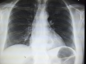 Bibasilar atelectasis tends to hamper the lung's ability to get the oxygen to the alveoli.
  25th ed. Mayo Clinic is a not-for-profit organization. General anesthesia is a common cause of atelectasis. Consolidation occurs when the normal, air-filled spaces of the lung are filled with the products of disease. David M. Hansell, David A. Lynch, H. Page McAdams, Alexander A. Bankier. These areas show increased density If it involves a whole lobe (lobar atelectasis), it may require further investigation; if it only affects a few A detailed Take a family member or friend with you to your appointment, if possible, to help you remember everything that is said. Your doctor may need to suction out excess mucus to allow you to take deep breaths and clear up your lungs. Home. For chest CT, the positive predictive value ranged from 1.5% to 30.7% and the negative predictive value ranged from 95.4% to 99.8%. Consult a doctor now! The peak is the top of the lung. 2.